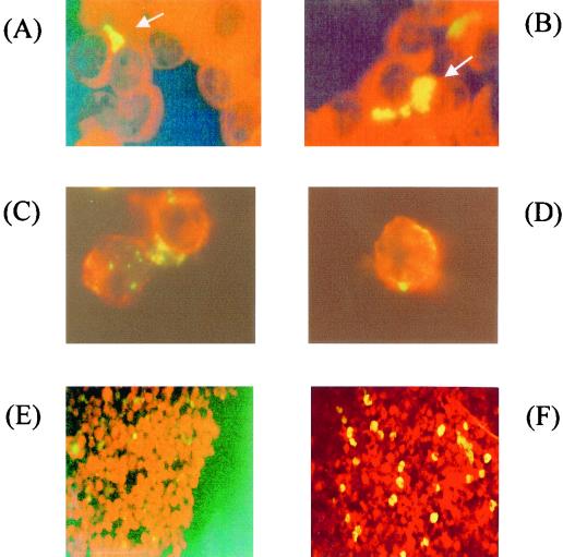
FIG. 1.
Fluorescence stain micrographs of C. pneumoniae-infected lymphocytes. (A and B) Human peripheral blood lymphocytes were infected with C. pneumoniae by gentle shaking and then incubated for 3 days at 37°C in 5% CO2. The infected cells were fixed on a glass slide by Cytospin and stained with FITC-conjugated anti-Chlamydia monoclonal antibody. Chlamydia inclusion bodies (arrow) were demonstrated in lymphocytes. Magnification, ×1,000. (C and D) Immunofluoresence micrographs of C. pneumoniae-infected mouse spleen lymphocytes. The mouse spleen lymphocytes were infected with C. pneumoniae by gentle shaking, incubated for 3 days, and then stained with PE-conjugated anti-mouse IgG CD3 antibody and FITC-conjugated anti-Chlamydia antibody after treatment with the permeabilization kit. Red color indicates surface molecules of lymphocytes. Green to yellow color indicates C. pneumoniae. Magnification, ×1,000. (E and F) Fluorescence micrographs of mouse T-cell-enriched lymphocyte cultures infected with C. pneumoniae. The purity of T cells in the cultures was more than 95% as determined by FACS analysis with anti-CD3 antibody. The T-cell cultures showed a few chlamydiae at 0 h (E). At 3 days after infection, there were many Chlamydia inclusion bodies (yellow spots) stained with FITC-conjugated anti-Chlamydia antibody in the culture (F). Magnification, ×200.

