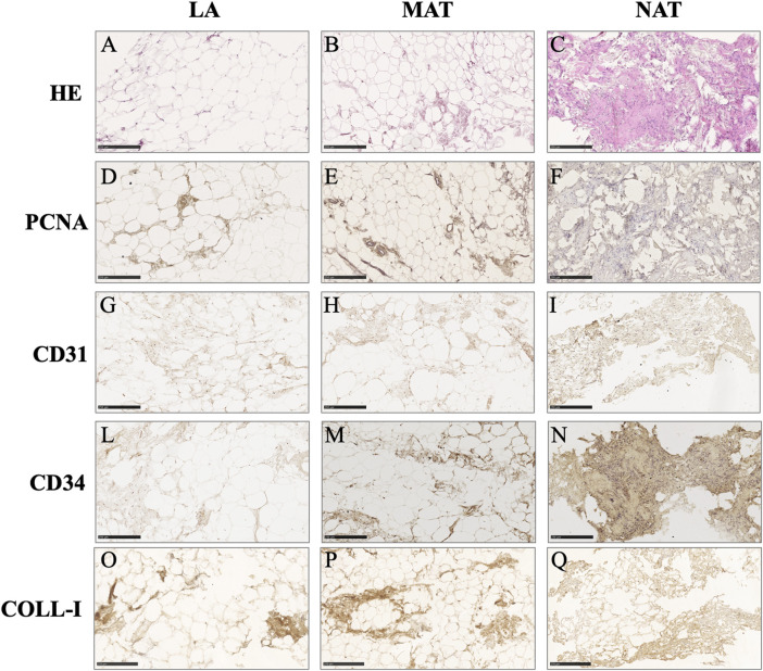
FIGURE 2.
Histochemical and immunohistochemical analysis of lipoaspirate (LA), micro-fragmented (MAT) and nanofat (NAT) formalin-fixed paraffin-embedded sections. Images are representative for each group. (A–C): hematoxylin-eosin (HE); (D–F): Proliferating cell nuclear antigen (PCNA); (G–I): CD31 (PECAM-1); (L–N): CD34; (O–Q): Collagen type I (COLL-I). Scale bars = 250 µm.

