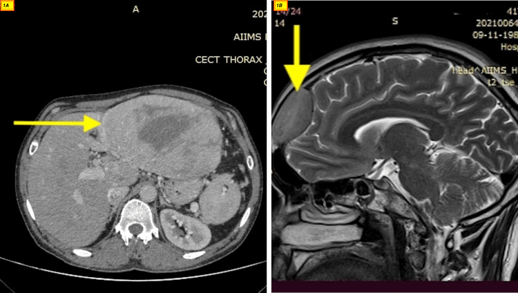
Figure 1. CT abdomen and MRI brain of the patient.
The image shows a large lobulated altered signal intensity lesion measuring 122x76x148 mm in the epigastric region with non-separate visualization of pancreas and left liver lobe (A). An active lesion in the right frontal region of the brain with lytic changes in the frontal bone (B).

