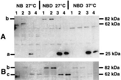
FIG. 2.
Immunoblot of yersiniae cells containing the respective constructs, developed with polyclonal rabbit anti-GFP antiserum (A) or polyclonal rabbit anti-luciferase antibody (B). Yersiniae were grown for 24 h under the conditions indicated at the top. Lane 1, WA-C(pYV08,pCJFY-GL); lane 2, WA-C(pYV08,pCJHE-GL); lane 3: WA-C(pYV08,pCJHE-G/FY-L); lane 4, WA-C(pYV08,pCJHE-L/FY-G). The GFP fusion protein (27 kDa) (a), GFP-LUC fusion protein (89 kDa) (b), and LUC fusion protein (62 kDa) (c) are indicated.

