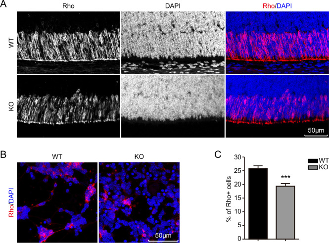
Figure 4.
Immunohistofluorescence staining of mouse retina and immunofluorescent staining of primarily cultured retina cells. (A) Retina rod cell genesis in P5.5 age mice of each genotype. Immunohistofluorescence staining of paraffin sections from P5.5 age mouse retina; rod cells stained with anti-Rho are shown in red colour, and the nucleus is presented in blue. (B, C) Immunofluorescent staining and statistical results of the primarily cultured cells from P5.5 age mouse retina. Rho: Rhodopsin, a marker of the rod cell. The errors bar presents a 95% CI. ***P<0.001.

