Figure 6.
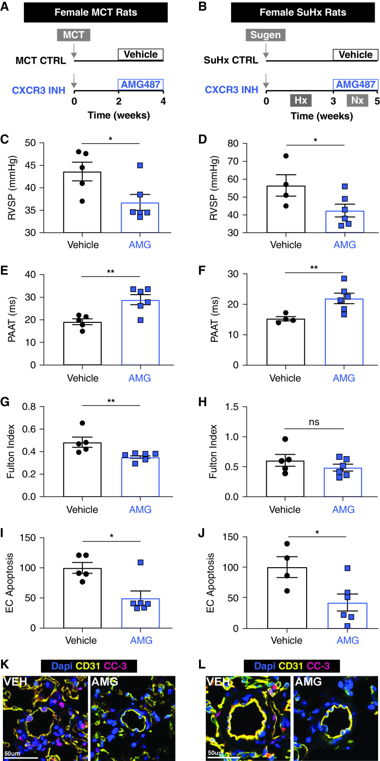
Blocking the activity of CXCL9 and CXCL10 is sufficient to rescue PH in two preclinical rat models. Schematic of in vivo experiments in female rats with (A) monocrotaline (MCT)-induced PH and (B) Sugen 5416-Hx (SuHx)–induced PH. All MCT model measurements are listed in the left column (A, C, E, G, I, and K), and SuHx measurements are listed in the right column (B, D, F, H, J, and L). (C and D) RVSP, (E and F) PAAT, and (G and H) Fulton index (RV/LV + IVS) measured in AMG487 (AMG)-treated PH rats compared with vehicle-treated PH controls. (I and J) Quantification and (K and L) representative images of apoptotic EC cells in CD31 (yellow) and cleaved caspase-3 (CC-3; pink) labeled sections from vehicle (VEH) and AMG-treated lungs. *P < 0.05 and **P < 0.01. Scale bars, 50 μm. CTRL = control; EC = endothelial control; INH = inhibition; IVS = intraventricular septum; LV = left ventricle; ns = not significant; PAAT = pulmonary artery acceleration time; RV = right ventricle; RVSP = right ventricular systolic pressure.

