Fig. 1.
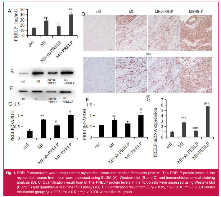
PRELP expression was upregulated in myocardial tissue and cardiac fibroblasts post MI. The PRELP protein levels in the myocardial tissues from mice were assessed using ELISA (A), Western blot (B and C) and immunohistochemical staining analysis (D). C. Quantification result from B. The PRELP protein levels in the fibroblasts were assessed using Western blot (E and F) and quantitative real-time PCR assays (G). F. Quantification result from E. *p < 0.05, **p < 0.01, ***p < 0.001 versus the control group; #p < 0.05; ##p < 0.01; ###p < 0.001 versus the MI group.

