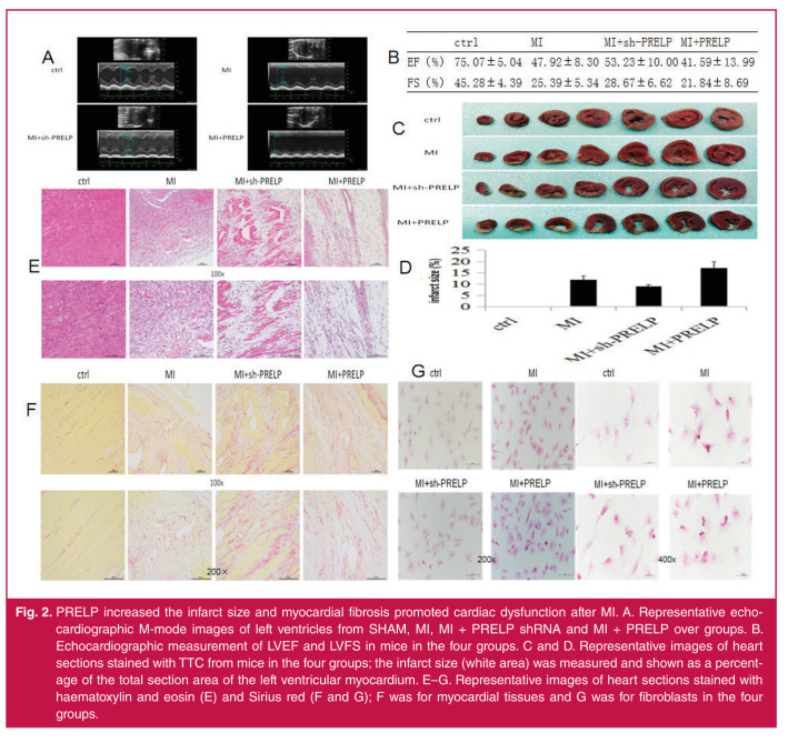
Fig. 2.
PRELP increased the infarct size and myocardial fibrosis promoted cardiac dysfunction after MI. A. Representative echocardiographic M-mode images of left ventricles from SHAM, MI, MI + PRELP shRNA and MI + PRELP over groups. B. Echocardiographic measurement of LVEF and LVFS in mice in the four groups. C and D. Representative images of heart sections stained with TTC from mice in the four groups; the infarct size (white area) was measured and shown as a percentage of the total section area of the left ventricular myocardium. E–G. Representative images of heart sections stained with haematoxylin and eosin (E) and Sirius red (F and G); F was for myocardial tissues and G was for fibroblasts in the four groups.

