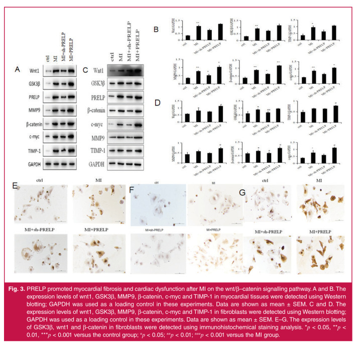
Fig. 3.
PRELP promoted myocardial fibrosis and cardiac dysfunction after MI on the wnt/β–catenin signalling pathway. A and B. The expression levels of wnt1, GSK3β, MMP9, β-catenin, c-myc and TIMP-1 in myocardial tissues were detected using Western blotting; GAPDH was used as a loading control in these experiments. Data are shown as mean ± SEM. C and D. The expression levels of wnt1, GSK3β, MMP9, β-catenin, c-myc and TIMP-1 in fibroblasts were detected using Western blotting; GAPDH was used as a loading control in these experiments. Data are shown as mean ± SEM. E–G. The expression levels of GSK3β, wnt1 and β-catenin in fibroblasts were detected using immunohistochemical staining analysis. *p < 0.05, **p < 0.01, ***p < 0.001 versus the control group; #p < 0.05; ##p < 0.01; ###p < 0.001 versus the MI group.

