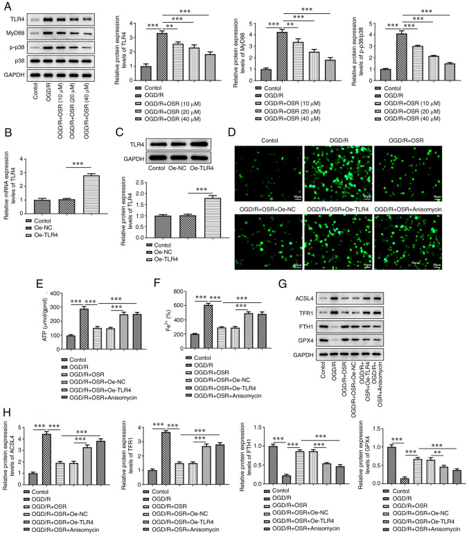
Figure 4.
OSR suppresses OGD/R-induced neuronal ferroptosis by inhibiting TLR4/p38MAPK signaling. (A) Protein expression levels of TLR4, MyD88, p-p38 and p38 were semi-quantified using western blotting. (B) mRNA and (C) protein expression levels of TLR4 in OGD/R-induced cells were assessed using reverse transcription-quantitative PCR and western blotting, respectively. The levels of (D) reactive oxygen species, (E) ATP and (F) Fe2+ were assessed in OGD/R-induced cells. Scale bar, 50 µm. (G) Protein expression levels of ACSL4, TFR1, FTH1 and GPX4 were (H) semi-quantified using western blotting. Data are presented as mean ± SD. Comparisons between multiple groups were performed using one-way ANOVA followed by Bonferroni's post hoc test for multiple comparisons. **P<0.01 and ***P<0.001. OGD/R, oxygen-glucose deprivation/reoxygenation; I/R, ischemia/reperfusion; p, phosphorylated; Oe, overexpression; NC, negative control; OSR, oxysophoridine; TLR, toll-like receptor; TFR1, transferrin 1; FTH1, ferritin 1; GPX4, glutathione peroxidase 4; ACSL4, acyl-CoA synthetase long-chain family member.

