
FIGURE 2.
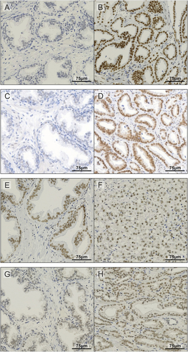
Representative images of IHC staining in prostate TMA. Examples of non-malignant (A) and prostate tumour tissue (B) stained for METTL3; non-malignant (C) and prostate tumour tissue (D) stained for METTL14; non-malignant (E) and prostate tumour tissue (F) stained for WTAP; and non-malignant (G) and prostate tumour tissue (H) stained for CBLL1.