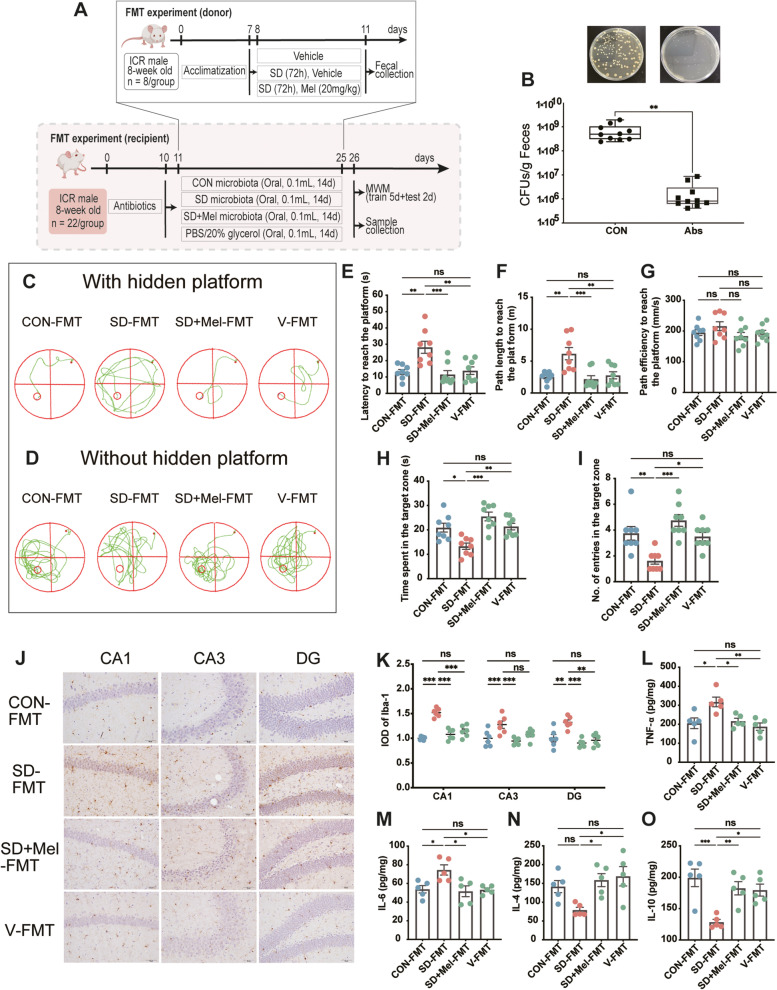
Fig. 2.
The gut microbiota mediated the neuroprotective effect of melatonin in memory impairment induced by sleep deprivation. A Schematic illustration of experimental design. B Comparison of bacterial colony-forming unit (CFU) in feces from control- and Abs-treated mice (n = 10). C Track plot of spatial memory test (with hidden platform). D Track plot of spatial memory test (without hidden platform). E Latency to reach the platform (n = 8). F Path length to reach the platform (n = 8). G Path efficiency to reach the platform (n = 8). H Time spent in the target zone (n = 8). I Number of entries into the target zone (n = 8). J Images of the immunohistochemical microglia in the different experimental groups. The immunohistochemical results were processed using ImageJ. Bar = 50 μm. K IOD of Iba1-positive cells in the hippocampal cornu ammonis (CA)1, CA3, and dentate gyrus (DG) regions (n = 6). L–O The levels of cytokines (TNF-α, IL-6, IL-4, and IL-10) in the hippocampus (n = 5). CON-FMT: receiving control microbiota FMT mice, SD-FMT: receiving sleep deprivation microbiota FMT mice, SD + Mel-FMT: receiving SD + Mel (20 mg/kg) microbiota FMT mice, V-FMT: receiving vehicle microbiota FMT mice. The data represent the mean ± SEM, p < 0.05 was set as the threshold for significance by one-way ANOVA followed by post hoc comparisons using Tukey’s test for multiple groups’ comparisons, *p < 0.05, **p < 0.01, ***p < 0.001

