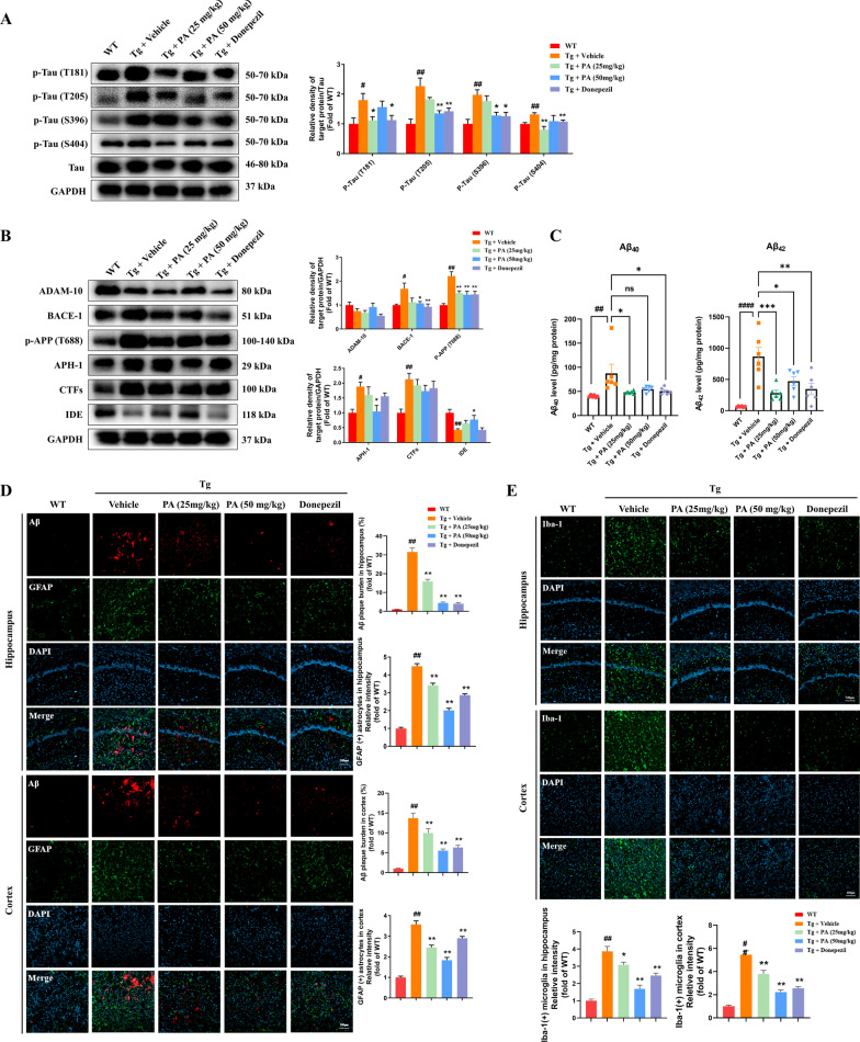
Fig. 2.
Effects of PA on the hyperphosphorylation of tau protein, Aβ pathology, GFAP-positive astrocytes and Iba-1-positive microglia in TgCRND8 mice. A Effects of PA on tau hyperphosphorylation at sites of T181, T205, S396 and S404 in TgCRND8 mice (n = 5–6); B effects of PA on the APP processing and APP phosphorylation in TgCRND8 mice. Representative western blotting images and quantitative analysis of the protein expressions of ADAM-10, BACE-1, p-APP (Thr 688), APH-1, CTFs and IDE (n = 5–6); C the levels of Aβ40 and Aβ42 (n = 6); D immunofluorescence staining of Aβ and GFAP in the hippocampus and cortex (n = 3); E immunofluorescence staining of Iba-1 in the hippocampus and cortex (n = 3). Data were expressed as the mean ± SEM (n = 3–6). #p < 0.05, ##p < 0.01 and ####p < 0.0001 compared with the WT group; *p < 0.05, **p < 0.01 and ***p < 0.001 compared with the Tg + vehicle group

