. 2023 Jan 24;307(2):e222379. doi: 10.1148/radiol.222379

Figure 3:

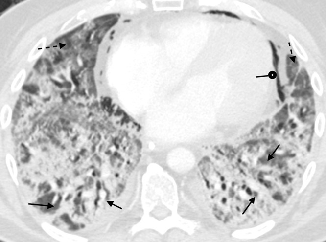
A 51-year-old man with COVID-19 infection. Axial contrast-enhanced CT image obtained 1 month after initial presentation. There is improved aeration of the dependent portions of the lungs; however, there is residual consolidation with contraction and architectural distortion (solid arrows). There is also persistent ground-glass opacity (dashed arrows) and new bronchial dilatation bilaterally. This appearance is suggestive of the organizing phase of acute lung injury. There is pneumomediastinum (round arrow) likely secondary to barotrauma from mechanical ventilation.

A 51-year-old man with COVID-19 infection. Axial contrast-enhanced CT image obtained 1 month after initial presentation. There is improved aeration of the dependent portions of the lungs; however, there is residual consolidation with contraction and architectural distortion (solid arrows). There is also persistent ground-glass opacity (dashed arrows) and new bronchial dilatation bilaterally. This appearance is suggestive of the organizing phase of acute lung injury. There is pneumomediastinum (round arrow) likely secondary to barotrauma from mechanical ventilation.