
Figure 6:
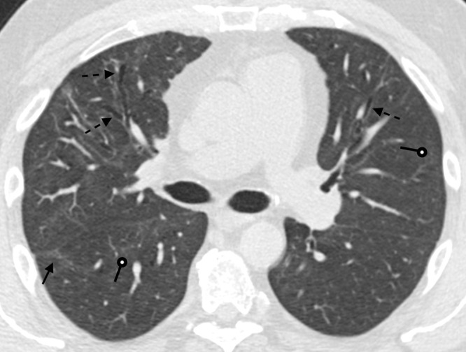
A 51-year-old man 9 months after COVID-19 infection. Axial contrast-enhanced CT image shows mild persistent anterior varicose bronchiectasis and architectural distortion best appreciated in the right middle lobe (dashed arrows). Faint patchy ground-glass opacities and reticulation are improved from 6-month imaging and barely perceptible (round arrows). Faint linear parenchymal bands have greatly improved (solid arrow). This may reflect the patient's new baseline.