. 2023 Jan 24;307(2):e222379. doi: 10.1148/radiol.222379

Figure 7:

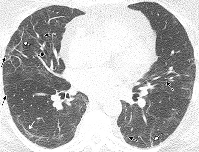
A 60-year-old woman with a history of hospitalization for moderate COVID-19 pneumonia requiring supplemental oxygen but not mechanical ventilation. Axial noncontrast CT image obtained 25 months after presentation for COVID-19 pneumonia shows bilateral thin parenchymal bands (white arrows), peripheral reticulation (black arrows), patchy ground-glass attenuation and reticulation, and traction bronchiectasis with architectural distortion (dashed black arrows). The fibrotic-like findings shown here appear to represent a new baseline given the 2-year period and have a pattern suggesting fibrotic sequelae of organizing lung injury in the setting of COVID-19. In other cases, parenchymal bands, ground-glass opacities, reticulation, and bronchial dilatation improve or resolve at follow-up imaging and cannot be interpreted as irreversible fibrosis without follow-up imaging.

A 60-year-old woman with a history of hospitalization for moderate COVID-19 pneumonia requiring supplemental oxygen but not mechanical ventilation. Axial noncontrast CT image obtained 25 months after presentation for COVID-19 pneumonia shows bilateral thin parenchymal bands (white arrows), peripheral reticulation (black arrows), patchy ground-glass attenuation and reticulation, and traction bronchiectasis with architectural distortion (dashed black arrows). The fibrotic-like findings shown here appear to represent a new baseline given the 2-year period and have a pattern suggesting fibrotic sequelae of organizing lung injury in the setting of COVID-19. In other cases, parenchymal bands, ground-glass opacities, reticulation, and bronchial dilatation improve or resolve at follow-up imaging and cannot be interpreted as irreversible fibrosis without follow-up imaging.