FIG 5.
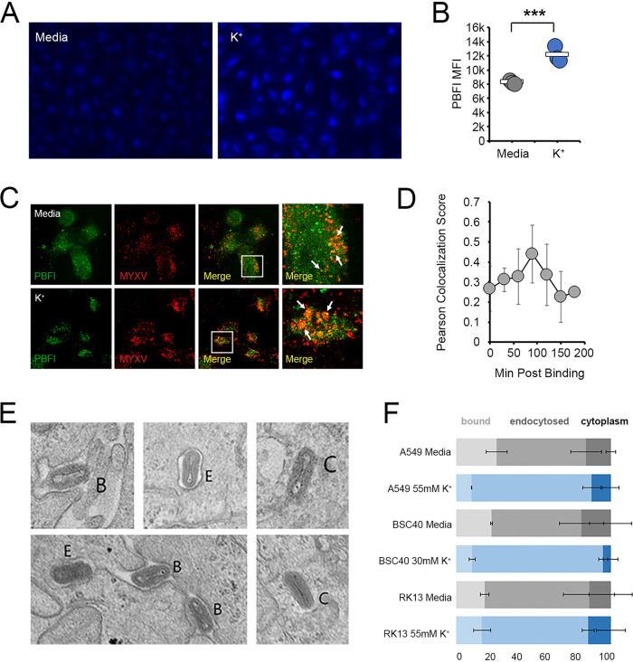
Elevated extracellular K+ inhibits the release of MYXV from the endosomes. (A and B) A549 cells were incubated in either regular medium (5 mM K+) (shown in gray) or medium containing 50 mM K+ (shown in blue) for 24 h. Cells were then stained with PBFI dye and analyzed for either the subcellular localization of K+ (A) or the intensity of PBFI fluorescence (B). (C and D) RK13 cells were incubated in either regular medium (5 mM K+) or medium containing 50 mM K+ for 24 h. Cells were then incubated with high numbers of MYXV virions containing an M094-Venus fusion protein (34) for the indicated times and then stained with PBFI dye. The colocalization of PBFI- and Venus-containing viral particles was then assayed using confocal microscopy. (C) Representative images of cells taken 90 min after virion adsorption. (D) Pearson colocalization scores of PBFI and Venus fluorescence obtained from images taken at the indicated time points. (E and F) Cells were incubated in either regular medium (5 mM K+) or medium containing 50 mM K+ for 24 h and then infected with MYXV at an MOI of 100. Thirty minutes after infection, cells were fixed and processed for electron microscopy. (E) Representative images showing viral virions that are bound at the cell surface (B), contained in membranous vesicles (E), or completely exposed in the cytoplasm (C). (F) Quantitation of the percentage of virions found at each subcellular location in the indicated cell types. The data shown in panel F represent data for more than 100 analyzed virions from each cell type from two independent experiments.

