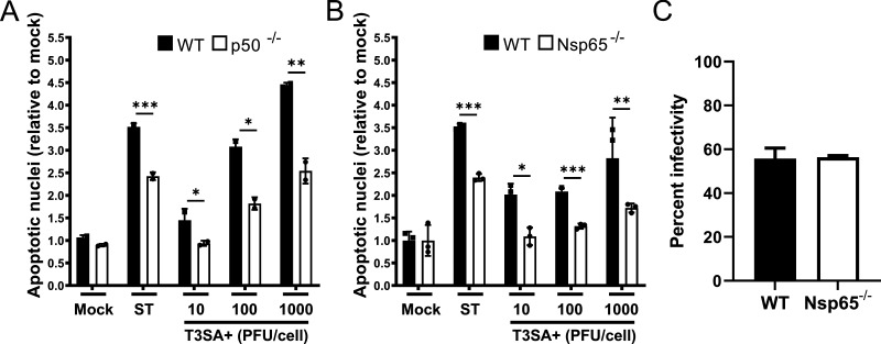
FIG 2.
Reovirus-induced apoptosis is diminished in cultures of cortical neurons that lack either NF-κB p50 or p65. Wild-type (WT), p50−/− (A), and Nsp65−/− (B) cortical neurons were adsorbed with reovirus T3SA+ ISVPs at the MOIs shown. At 24 h postadsorption, the percentage of apoptotic nuclei was quantified following AO staining. Staurosporine (ST, 10 mM) was used as positive control. Each point is expressed as the mean percentage of apoptotic cells for triplicate wells. (C) The percentage of infected WT and Nsp65−/− cortical neurons was quantified 24 h postadsorption with 1,000 PFU/cell T3SA+. Error bars indicate the SD. *, P < 0.05; **, P < 0.01; ***, P < 0.001 (as determined by Student t test compared to results for p50−/− or Nsp65−/− neurons at the same MOI).

