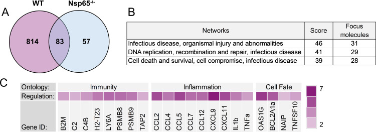
FIG 5.
Expression of genes influenced by reovirus infection and NF-κB p65 in the brain. Wild-type (WT) and Nsp65−/− newborn mice were inoculated intracranially with 10 PFU of reovirus T3SA+ or PBS. Cortices were removed at 6 days postinoculation and processed for RNA purification and mRNA sequencing. RNA samples prepared from infected mice were matched for viral titer. (A) Venn diagram of differential expression analysis using DeSeq2 package in R between WT infected versus mock infected (pink) and Nsp65−/− infected versus mock infected (blue). (B) Networks with the most differentially expressed genes were identified using Ingenuity Pathway Analysis. (C) Log2-fold change heat map indicating the expression levels of selected genes. The log2-fold change was calculated based on the DESeq2 analysis of RNA levels in WT-infected brains relative to Nsp65−/−-infected brains.

