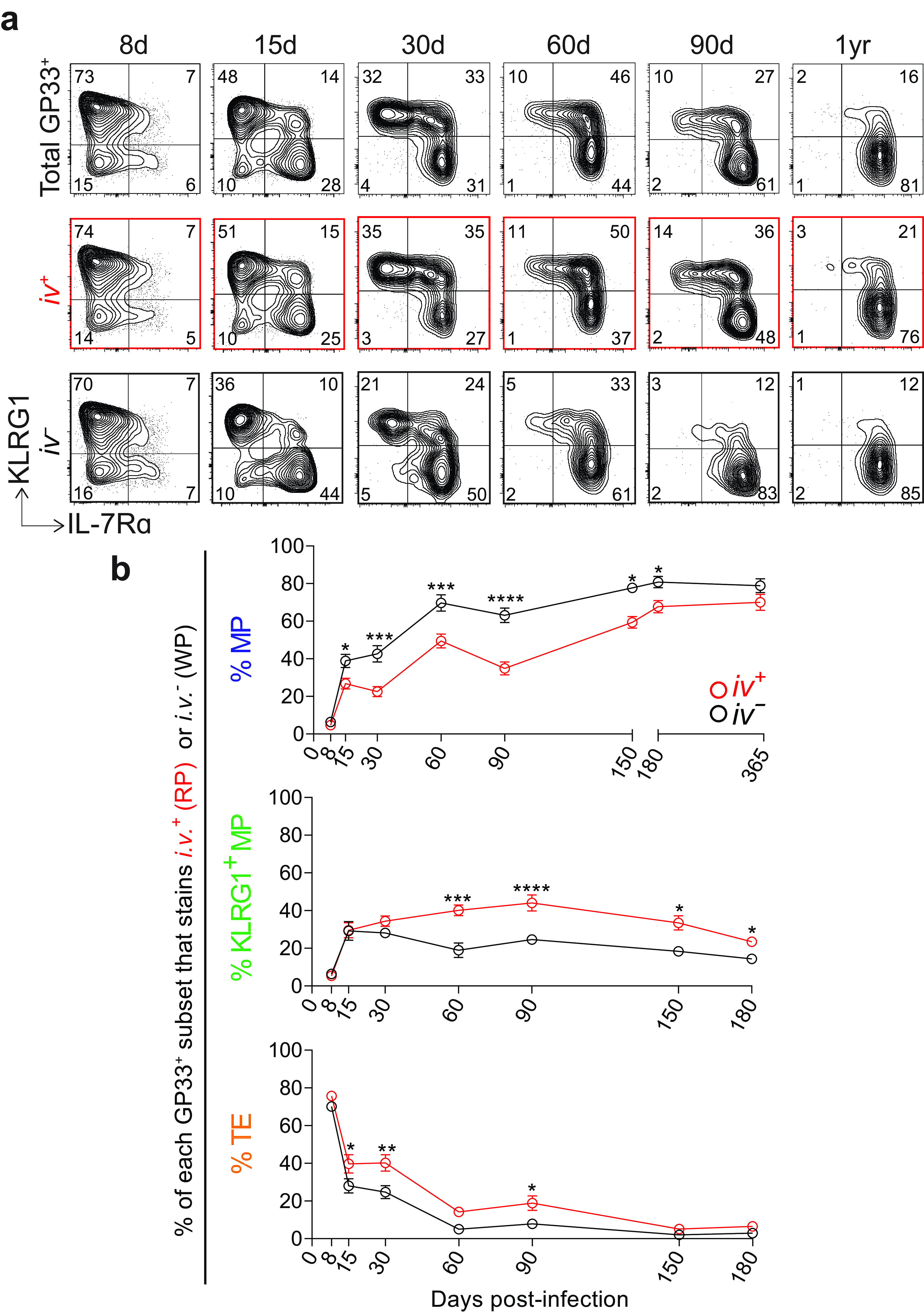
FIG 7.

MPs localize to the splenic white pulp as early as 1 to 2 weeks after acute infection. (a) Representative flow plots showing KLRG1 and IL-7Rα expression on total GP33+ CD8 T cells in the spleen (top row). Below, GP33+ CD8 T cells are further gated by i.v. labeling, either i.v.+ (RP) (middle row) or i.v.− (WP) (bottom row). (b) Summary of the frequency of each memory subset within GP33+ CD8 T cells in the RP or WP. Shown are the mean and SEM; n = 5 to 9 mice per time point. *, P ≤ 0.05; **, P ≤ 0.01; ***, P ≤ 0.001; ****, P ≤ 0.0001 (all determined using two-way ANOVA with Sidak’s test for multiple comparisons).
