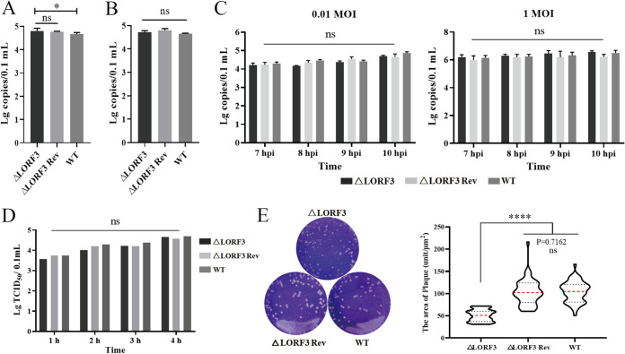
FIG 6.
LORF3 in the viral adsorption, invasion, replication, and release processes and cell-to-cell spread. (A) DEF cells were infected with the indicated viruses at 4°C, and cell samples were collected after 2 hpi to detect the copies and analyze the adsorption efficiency of viruses. (B) After viral adsorption was completed, the cells were incubated at 37°C for 1 hpi, and the viruses in the cell were quantified to analyze the viral invasion efficiency. (C) qPCR assays for viral genome copies in cells infected with low- and high-dose DPV in consecutive periods. (D) Cells were replaced with fresh maintenance solution after DPV infection, the supernatant was collected every 1 hpi, and the release of mature virions was detected by virus titer. (E) Crystal violet assay to test the cell-to-cell spread. Representative plaques showing 50 plaques per sample were measured to quantify the results at the right. Plates were scanned, and plaque diameters were measured in Image J (ns, P > 0.05; *, P < 0.05; ****, P < 0.0001).

