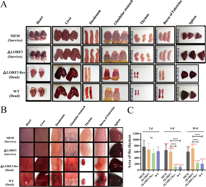
FIG 8.
Autopsy histopathology of designated viruses-infected ducks. (A) Histopathological damage records of the dead ducks infected with revertant and parental strains and the surviving ducks infected with the deletion strain at 6 dpi. (B) Magnified view of the local lesion of the organs in panel A. (C) Thymus atrophy due to DPV infection. Each group's thymus area of ducks was scanned and measured with ImageJ within 10 days of the attack. Results are shown based on the thymus area of ducks in the control group as 100% (****, P < 0.0001).

