
FIG 2.
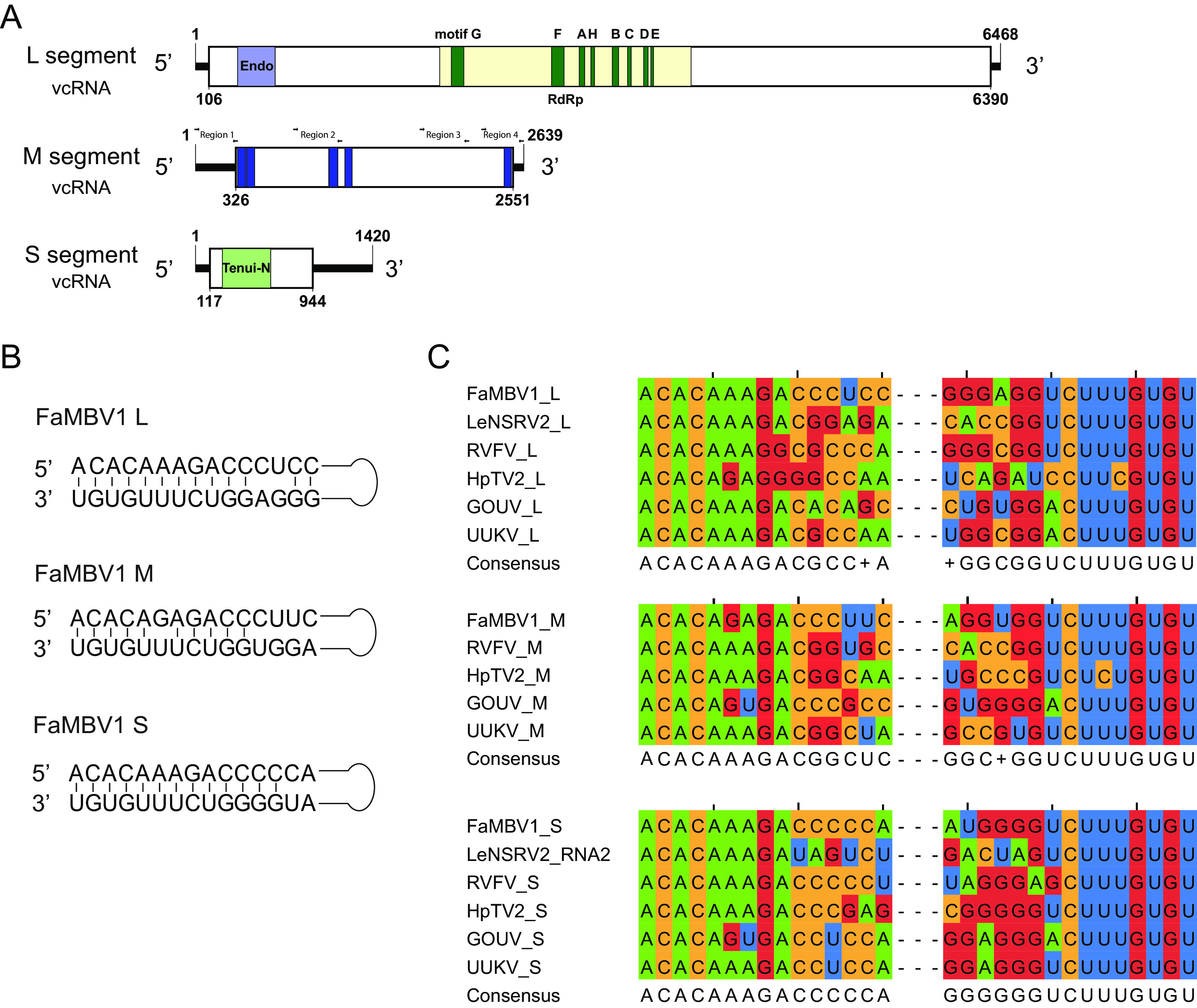
Schematic diagram of the FaMBV1 genome and features. (A) Schematic diagram of the FaMBV1 genome. vRNA, viral RNA strand; vcRNA, viral RNA complementary strand. Open reading frames are represented by rectangles. Conserved domains are presented in an acid green box. Four different regions with primers used for M segment detection by PCR are shown. (B) Panhandle structures are formed by the 5′ and 3′ termini of FaMBV1 L, M, and S segments. (C) Comparison of FaMBV1 L, M, and S segment terminal nucleotide sequences with other bunyaviruses; consensus sequences are presented. LeNSRV2, Lentinula edodes negative-stranded RNA virus 2; RVFV, Rift Valley fever virus; HpTV2, Huángpí tick virus 2; GOUV, Gouléako virus; UUKV, Uukuniemi virus.