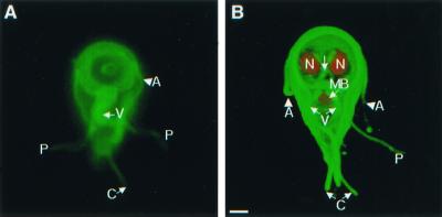
FIG. 1.
Important cytoskeletal structures of nondividing Giardia trophozoites demonstrated by video microscopy (A) or confocal microscopy (B). (A) Ventral view of a giardia labeled green on its surface with Alexa Fluor 488, with highlighted anterior (A), posterolateral (P), caudal (C), and ventral (V) flagella. (B) A three-dimensional confocal micrograph of the ventral surface of another giardia. Its surface was labeled green with Alexa Fluor 488, the nuclei (N) were stained red with propidium iodide, and the median body (MB) was stained red with monoclonal antibodies to bovine tubulin. Bar, 2 μm.

