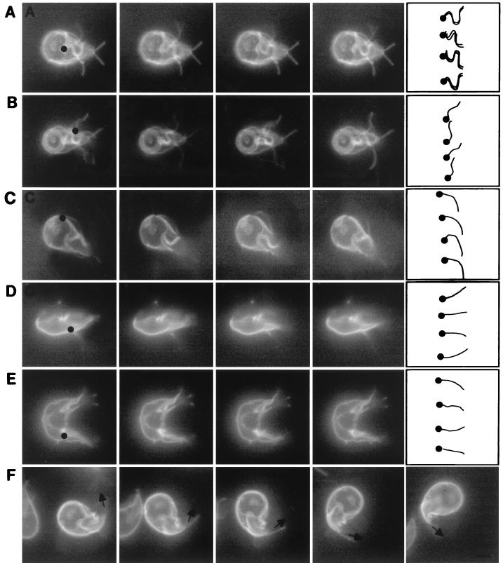
FIG. 2.
Videos of adherent and swimming giardia, each of which was labeled on its surface with Alexa Fluor 584. (A) Ventral flagella of an adherent giardia moved with a series of bends, which originated where the ventral flagella exited the cytosol (black dot) and propagated to the tip (drawing at right of row A). In contrast, the anterior, posterolateral, and caudal flagella of this adherent giardia were still. (B) The posterolateral flagellum of a second adherent giardia moved with a series of bends which originated where the posterolateral flagella exited the cytosol (black dot) and propagated to the tip (drawing at right of row B). The beat of the posterolateral flagella had the same frequency and wavelength but less amplitude than that of the ventral flagella. (C) An anterior flagellum of a swimming giardia moved with a single bend of low amplitude (drawing at right of row C), which originated where the anterior flagellum exited the adherence disc (black dot). More subtly, the beating motion of the caudal flagella in a plane perpendicular to the adherence disc caused the tails of swimming giardia to go in and out of focus. In contrast, the wavelike bending of the caudal flagella (drawings in rows D and E) was easier to see when swimming (D) or dividing (E) giardia were viewed in profile. The bending of the caudal flagella begins within the cytosol of the tail (black dots in rows D and E). (F) The tail of a turning giardia remained curved in the direction of the turn, like the rudder on a boat. Bars in the drawing at the right of row F indicate direction of the caudal flagella. Each set of videos was composed of consecutive frames shot at 20 frames per s. Bar, 5 μm.

