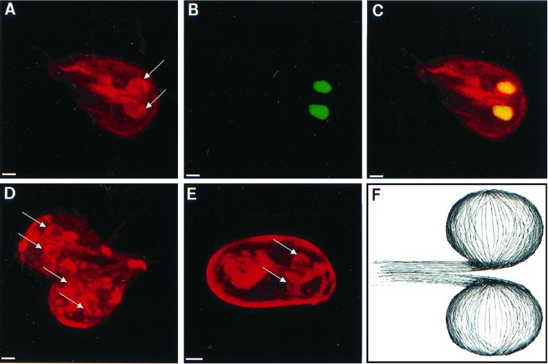
FIG. 6.
Three-dimensional confocal micrographs highlighting microtubules which form perinuclear tethers. (A to C) Micrographs of a giardia which was stained with polyclonal antibodies to α-tubulin (red in panels A and C) and Sytox green (green in panel B and yellow in panel C) show tethers of microtubules (arrows) that surround both nuclei. A dividing giardia (D) and an encysted giardia (E) stained with the same antitubulin antibodies have prominent perinuclear tethers (arrows). Note that the wall of the cyst is nonspecifically stained with the antibodies. Bars, 2 μm. (F) Cartoon of perinuclear tethers of microtubules which connect to microtubules in the central axis of the parasite.

