FIG. 7.
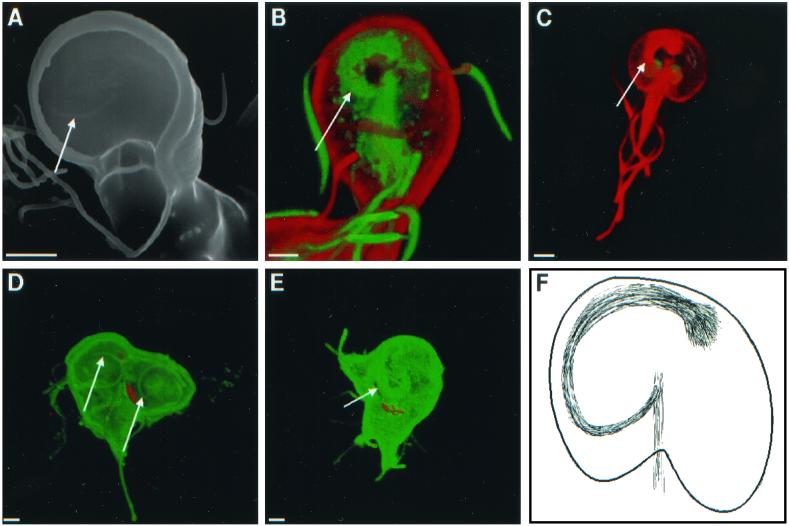
Scanning (A) and confocal (B to E) micrographs highlight spirals of microtubules (arrows), which underlie adherence discs. (B) Giardia labeled red on its surface with Alexa Fluor 584 and stained green with antibodies to acetylated tubulin. (C) Giardia stained red with antibodies to acetylated tubulin. (D and E) Dividing giardia labeled green on their surface with Alexa Fluor 488 and with median bodies labeled red with monoclonal antibodies to bovine tubulin. Bars, 2 μm. (F) Cartoon shows the spiral of microtubules, which underlies the disc and attaches to the axis of microtubules.

