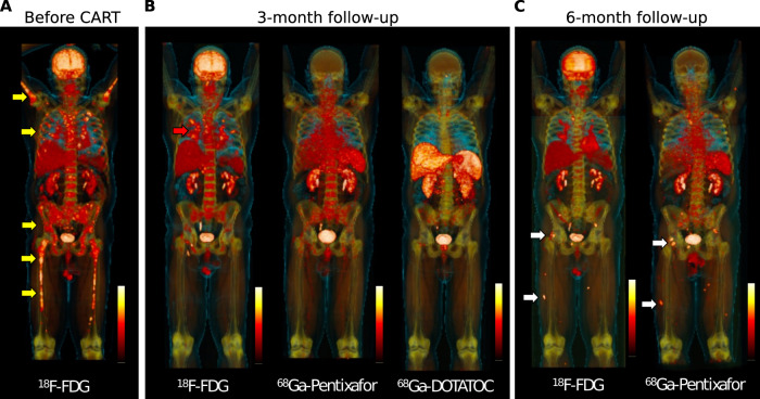
Fig. 1. Whole body PET imaging at baseline and during follow-up using multiple PET tracers.
A FDG-PET at baseline shows multiple focal lesions at the axial and appendicular skeleton (yellow arrows). B Follow-up assessment at 3-month after CAR T infusion shows full resolution of focal lesions, but residual FDG uptake located to the lung (red arrow). Matched-simultaneous CXCR4-targeted Pentixafor and DOTATOC PET did not show focal uptake. C Matched-simultaneous FDG and CXCR4 PET 6 months after CAR T depicted new focal lesions at the pelvis and lower limbs in line with relapse (white arrows).

