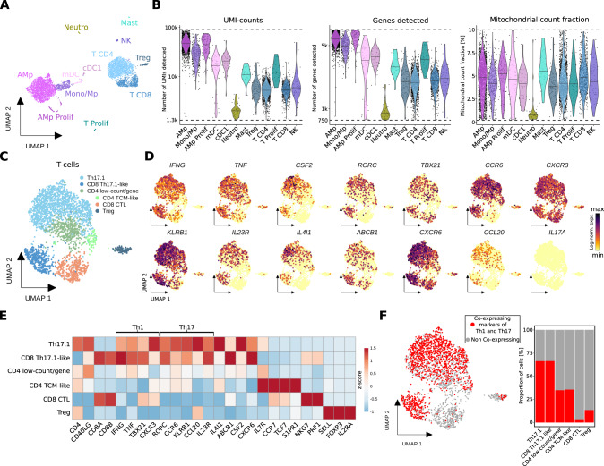
Fig. 2. Single-cell analysis of bronchoalveolar lavage of a MM patient with FDG-avid pulmonary manifestation on PET-imaging suggests Th17.1-driven autoimmune phenomenon following anti-BCMA CAR T treatment.
A UMAP embedding of 6,042 single-cell transcriptomes from two technical replicates from BAL (bronchoalveolar lavage) of a MM patient at three months follow-up after CAR T therapy (CART-BAL). Cell type annotation was based on expression of canonical marker genes (Supplementary Fig. 2D). B Data quality metrics across cell types in CART-BAL depicted in violin plots. Dashed lines represent thresholds that were used for quality control filtering. For UMI-counts and genes detected, log10-scale is shown. Lines in violins show medians per cell type. C UMAP embedding of 2,778 T-cells (T CD4, T CD8, Treg from A) from CART-BAL colored by subset annotation. D Log-normalized gene expression of Th1-associated genes (IFNG, TNF, TBX21, CXCR3), Th17-associated genes (RORC, CCR6, KLRB1, IL23R, CCL20) and further selected genes (CSF2, IL4I1, ABCB1, CXCR6) that together characterize Th17.1-cells and IL17A color-coded and projected onto the UMAP embedding from C. E Heatmap showing the z-score of mean log-normalized expression of selected genes per T-cell subset identified in C. F Co-expression of Th1-associated (IFNG, TNF, TBX21, CXCR3) and Th17-associated (CCL20, IL23R, RORC, CCR6) genes in individual cells was assessed and projected onto the UMAP embedding as well as depicted as bar plot. Cells were deemed co-expressing when at least one Th1- and one Th17-associated gene was detected. AMp alveolar macrophage, Mono monocyte, Mp macrophage, CTL cytotoxic T-lymphocyte, TCM central memory T-cell.

