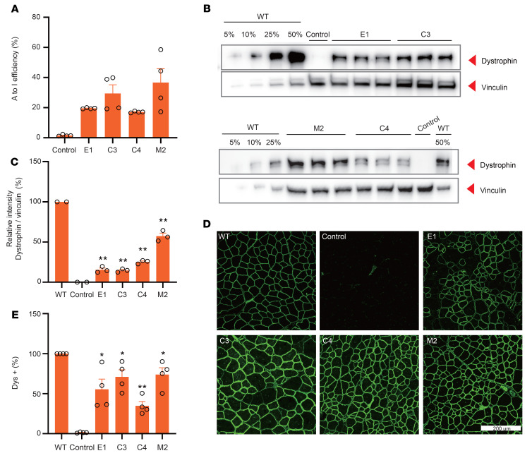
Figure 4. AAV-mxABE robustly rescues dystrophin expression in TA 6 weeks after injection.
(A) Deep sequencing of in vivo RNA editing 6 weeks after i.m. injection with AAV9-E1, -C3, -C4, and -M2 constructs (n = 4). (B) Western blot analysis of dystrophin (MilliporeSigma, D8168) protein expression in TA muscles of WT and DMDE30mut mice. Intramuscular injection of saline in the DMDE30mut mice was the control. Vinculin was used as the loading control. (C) Quantification of dystrophin expression from Western blots after normalization to vinculin (n = 3). (D) Immunohistochemistry of dystrophin in TA muscles 6 weeks after i.m. injection with different AAV9 constructs. Dystrophin (Abcam, ab15277) is indicated in green. Scale bar: 200 μm. (E) Quantification of Dys+ fibers in cross sections of TA muscles (n = 4). Dots and bars represent biological replicates and are mean ± SEM. Unpaired 2-tailed Student’s t test. *P < 0.05, **P < 0.01 vs. control.

