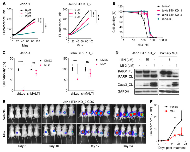
Figure 3. MALT1 inhibition by MI-2 decreases MALT1 paracaspase activity and suppresses proliferation in MCL cells.
(A) Endogenous MALT1 cleavage activity detected in JeKo-1 cells and JeKo BTK KD-2 cells upon MALT1 inhibition by MI-2 at the indicated concentrations and treatment times. Each treatment for the indicated cell lines was set up in triplicate. Statistical significance was determined based on the F test of the slope from the linear regression model and multiple comparison was adjusted using Šídák’s approach. (B) MALT1 inhibitor MI-2 potently inhibited viability in JeKo-1, JeKo-R, and JeKo BTK KD-1 and -2 cells. (C) MI-2 effectively inhibited viability in JeKo-1 cells and JeKo BTK KD-2 cells, but not their counterparts with stable MALT1 KD. Error bars were generated from at least 3 independent replicates (B and C). (D) MI-2 induced cleavage (CL) of full-length PARP (PARP_FL) and caspase 3 in JeKo BTK KD-2 cells and primary patient cells. (E and F) NSG mice bearing luciferase-expressing JeKo BTK KD-2–derived subcutaneous xenografts were treated with vehicle (n = 5) or MI-2 (n = 5) at 25 mg/kg daily via intraperitoneal injection for 24 days. Tumor growth was monitored by live animal luminescence imaging (E) and the luciferase flux was plotted (F). Two-way ANOVA was used in C and F, and statistical significance was determined based on the adjusted P values using Šídák’s method. *P < 0.05; ***P < 0.001; ****P < 0.0001.

