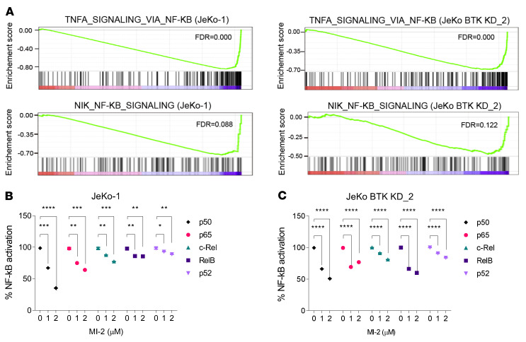
Figure 4. MALT1 inhibition by MI-2 suppresses NF-κB signaling in MCL cells.
(A) The enrichment score of TNF-α signaling via NF-κB (upper panels) and NF-κB–inducing kinase (NIK) NF-κB signaling (bottom panels) in JeKo-1 (left panels) and JeKo BTK KD-2 (right panels) cells. (B and C) Activity of all 5 NF-κB family members was reduced upon MALT1 inhibition by MI-2 in JeKo-1 (B) and JeKo BTK KD-2 (C) cells. Error bars were generated from 3 independent replicates (B and C). Two-way ANOVA was used in B and C, and statistical significance was determined based on the adjusted P values using Šídák’s method. Data represent mean ± SD. *P < 0.05, **P < 0.01, ***P < 0.001, ****P < 0.0001.

