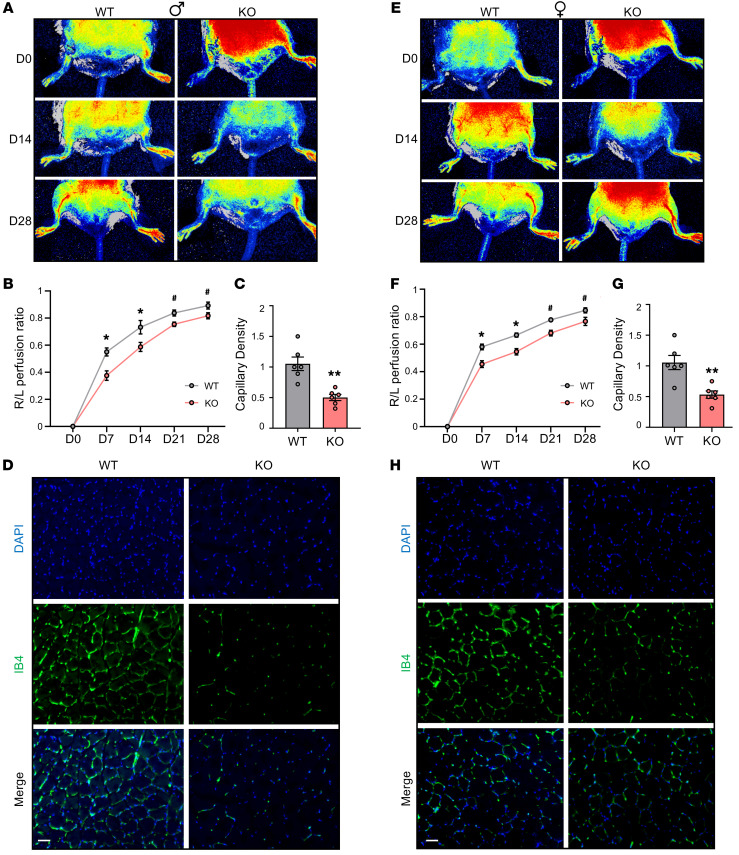
Figure 4. Leene-KO mice have impaired hind limb blood flow recovery after arterial ligation.
Male (A–D) and female (E–H) mice were fed an HFHS diet for 16 weeks, followed by femoral artery ligation on the right hind limb and sham operation on the left on day 0 (D0). Perfusion recovery rate was measured at various time points (D0, D7, D14, D21, and D28) after femoral artery ligation by laser speckle flowgraphy. Data show blood perfusion ratio of the right to left (R/L) hind limb. Representative images (A and E) and quantitative analysis (B and F) (n = 8–10/group). (C and G) Quantification of capillary density based on IB4 staining (n = 6/group) and (D and H) representative images of IB4 (green) and DAPI (blue) staining in the gastrocnemius muscle collected 7 days after HLI. Scale bars: 50 μm. Data are represented as mean ± SEM. #P = 0.05, *P < 0.05; **P < 0.01 based on 2-tailed Student’s t test.

