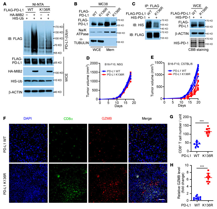
Figure 5. MIB2 catalyzes PD-L1 ubiquitination on K136 residue.
(A) In vivo PD-L1 ubiquitination by MIB2 in 293T cells transfected with PD-L1 WT or the K136R mutant expression construct. (B) Immunoblotting (IB) analysis of PD-L1 protein levels in whole-cell extract (WCE) and membrane fractions (Mem) from MC38 cells expressing WT PD-L1 or the K136R mutant. (C) Immunoprecipitation (IP) and immunoblotting (IB) analysis of A375 cells with WT PD-L1 or K136R mutant interacting with purified HIS–PD-1. (D and E) Tumor growth curves of (D) NSG and (E) C57BL/6 mice inoculated with B16-F10 cells expressing PD-L1 WT or the K136R mutant (n = 5 per group). (F–H) CD8 and granzyme B (GZMB) immunostaining in the B16-F10 PD-L1 WT and PD-L1 K136R mutant tumors from C57BL/6 mice (n = 9); 3 tissue slides per tumor. (F) Representative images. Scale bar: 50 μm. (G) Quantification of CD8+ T cells. (H) Relative GZMB level. Unit = 262,144 μm2 (the area of the tumor tissue). **P < 0.01; ***P < 0.001, by unpaired, 2-tailed t test between 2 groups (G and H). Data are shown as the mean ± SEM.

