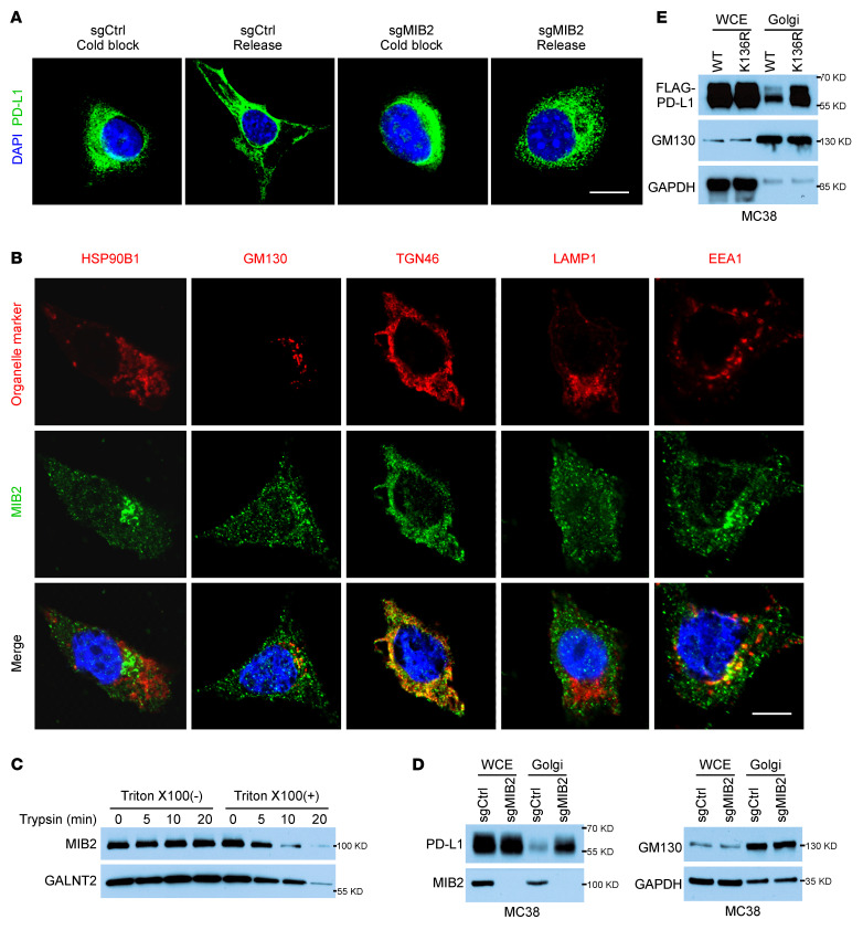
Figure 6. Ubiquitination by MIB2 is required for PD-L1 exocytosis.
(A) Immunofluorescence analysis of PD-L1 in B16-F10 cells after cold block release. Scale bar: 10 μm. (B) Colocalization of MIB2 and subcellular organelles markers in B16-F10 cells. Scale bar: 10 μm. (C) Immunoblotting (IB) analysis of MIB2 and Galnt2 in trypsin-digested Golgi fractions with or without permeabilization. (D) IB analysis of PD-L1 in the whole-cell extract (WCE) and isolated Golgi from MC38 cells. (E) IB analysis of PD-L1 in the WCE and isolated Golgi from MC38 cells expressing PD-L1 WT or K136R mutant.

