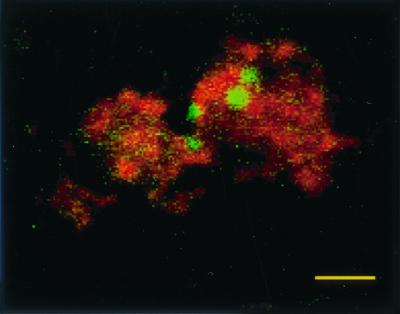
FIG. 3.

Confocal laser micrograph of Salmonella serovar Pullorum 449/87(pBRD940) expressing GFP within splenic macrophages counterstained with wheat germ agglutinin–Texas Red-X. Macrophages were isolated from infected birds 21 days following infection. Bar = 10 μm.
