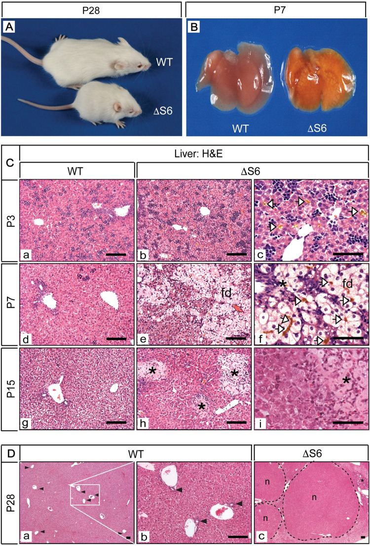
Fig 1. Perinatal deletion of hepatic Rps6 retards growth and results in cholestatic liver disease.
(A) A WT and runted ΔS6 mouse at postnatal day 28 showing that hepatic Rps6- deficiency retards neonatal growth. (B) Gross appearance of livers from a WT and a ΔS6 mouse at post-natal day 7 (P7). Severe yellowing of the ΔS6 liver is indicative of cholestatic disease. (C) Photomicrographs of H&E stained sections of liver from WT mice at P3, P7 and P15 (a, d and g) and age-matched ΔS6 mice (b-i). Canalicular accumulation of bile (yellow deposits, open arrowheads) is evident in ΔS6 livers at P3 and P7 and feathery degeneration (fd) of hepatocytes is evident at P7. At P15, bile infarcts (*) resulting from bile leakage due to canalicular or hepatocyte membrane rupture can be seen throughout the parenchyma of ΔS6 livers (Original magnifications, a, b, d, e, g, h (x 125; 50μ scale bars); c, f and i (x 375; 25μ scale bars). (D) Photomicrographs of H&E stained sections of liver from a WT mouse (a and b) and a ΔS6 littermate (c) at 4 weeks of age. In contrast to the WT liver (a and b) which shows an abundance of bile ducts (arrowheads), ΔS6 livers appear to either lack or have a paucity of bile ducts (c) while nodules (n) and areas of biliary hyperplasia (*) are prominent indicating that Rps6 insufficiency has severely disrupted liver architecture. (Original magnifications, a and c (x 32.5); b (x 125)). Scale bars; all 50μ.

