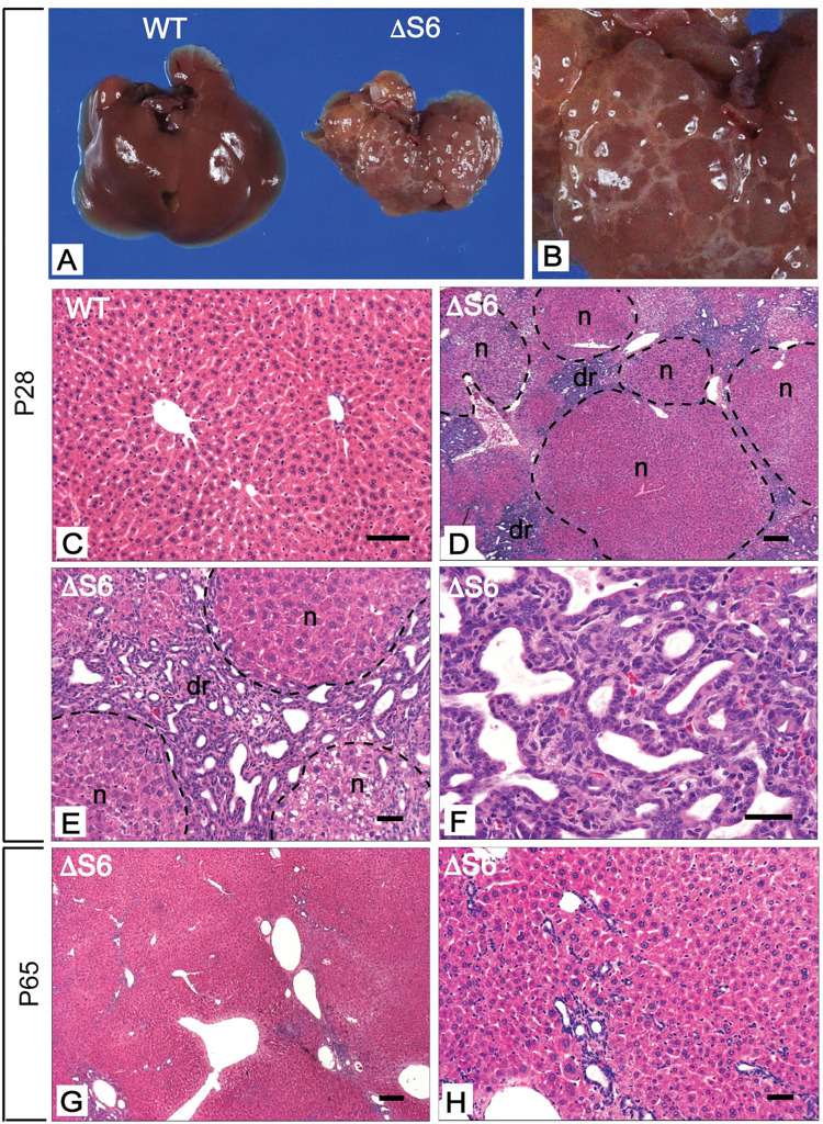
Fig 3. Perinatal deletion of hepatic Rps6 results in hypoplastic livers and triggers regeneration.
(A) Gross appearance of livers from a WT and ΔS6 mouse at postnatal day 28 (P28). The ΔS6 liver is smaller than the WT liver, is discolored and has an uneven mottled appearance. (B) Close-up image of ΔS6 liver in a) highlighting “cystic-like” nodules on the surface of the liver. (C-H) Photomicrographs of H&E stained sections of liver from a WT mouse (C) and ΔS6 mice at P28 (D-F) and P65 (G and H). At P28, ΔS6 livers display an abundance of regenerative nodules (n) and a prominent ductular reaction (dr) signifying dynamic regeneration in response to severe injury. By P65, the absence of regenerative nodules indicates that the regenerative response has largely dissipated. However, hepatocyte morphology is heterogeneous and remnants of the dr persist as seen by the presence of irregular luminal structures in the vicinity of portal veins (g, h). (Original magnifications, C (x 112); D, G (x 32); E, H (x 125) and F (x 250)). Scale bars correspond to 50μ for C, E, F and H and 200μ for D and G.

