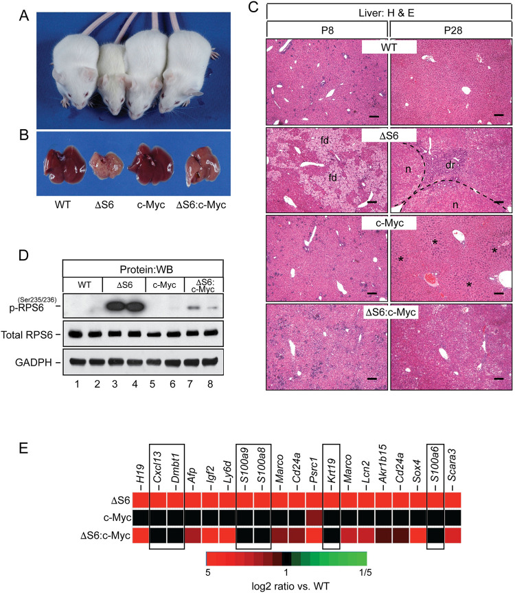
Fig 7. Overexpression of c-Myc rescues the growth defect and eliminates the requirement for ΔS6 livers to regenerate by preserving hepatocyte viability.
(A) Picture of a 28 day old WT, ΔS6, c-Myc and ΔS6:c-Myc mouse. The ΔS6:c-Myc mouse (far right) is indistinguishable from the WT mouse (far left) in terms of size and lacks the jaundiced (yellowed) coat of the ΔS6 mouse (second from left). (B) Gross appearance of livers from 28 day old WT, ΔS6, c-Myc and ΔS6:c-Myc mice. Note the smooth and less jaundiced appearance of the ΔS6:c-Myc liver relative to the ΔS6 liver. (C) Photomicrographs of H&E stained liver sections of 8 and 28 day old WT, ΔS6, c-Myc and ΔS6:c-Myc mice showing the absence of feathery degeneration at P8 and lack of regenerative nodules or a dr at P28 in livers of ΔS6:c-Myc compared to ΔS6 mice. Dotted lines depict nodule boundaries. Asterisks (*) denote characteristic regions of hepatocyte hypertrophy in Alb-c-Myc livers (Original magnifications, x 62.5; scale bars 50μ). (D) Western blot showing that mTOR-dependent phosphorylation of RPS6 is suppressed by overexpression of c-Myc in ΔS6:c-Myc livers. GAPDH; protein load control. (E) Heat map showing that overexpression of c-Myc normalizes the innate immunity molecular signature comprising NF-κB target genes and DAMPs (boxed areas), but not imprinted genes, in ΔS6 livers.

