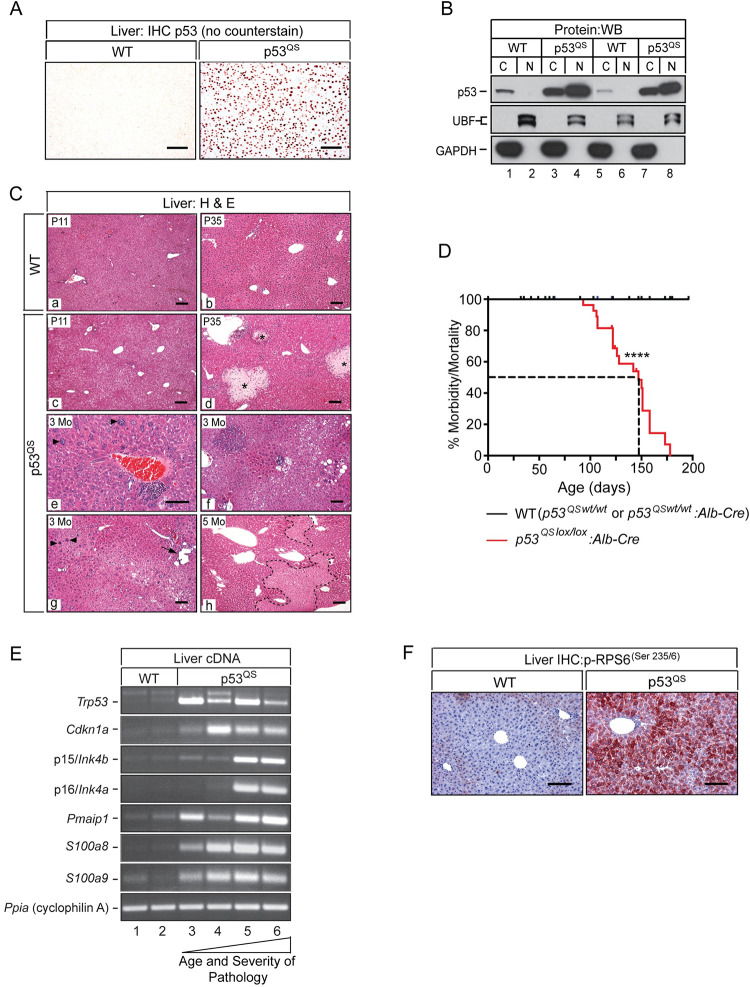
Fig 8. Hepatoblast-specific expression of p53QS results in chronic liver failure preceded by induction of senescence and innate immunity and activation of mTOR.
(A) p53 IHC performed on liver sections from a WT (left) and p53QS expressing mouse (right) showing robust nuclear expression of the p53QS mutant in hepatocytes. Original magnifications, x125. (AEC Chromogen (orange/red), no counterstain). (B) Western blot of fractionated cytoplasmic (C) and nuclear (N) proteins isolated from the livers of 2 WT mice (lanes 1, 2, 5 and 6) and 2 p53QS mice (lanes 3, 4, 7 and 8) with a p53-specific antibody. Immunoblotting of the same lysates using antibodies specific for the nuclear protein UBF (upstream binding factor) and the cytoplasmic protein GADPH confirm enrichment of nuclear and cytoplasmic proteins after fractionation. (C) H&E stained sections of WT (a, b) and p53QS livers (c-h) showing age-dependent progression of disease in p53QS livers. Regional hepatocyte vacuolization, evident at P11 (c) is followed by focal hepatocyte necrosis (biliary infarcts, *) by ~5 weeks of age (d). After ~3 months, hepatocytes show increasing heterogeneity across the lobule (e, f, g). Karyomegaly (enlarged nuclei (arrowheads, e and g)), regional hepatocyte vacuolization and biliary dilatation (g, arrow) are common. By ~5 months of age, p53QS livers show evidence of widespread parenchymal loss (h, necrotic areas bounded by dashed lines) signifying ongoing liver decompensation in the absence of regeneration. Original magnifications; a, b, c, d, g, h and f, x62.5; e, x125. (D) Kaplan-Meier curve of morbidity and mortality in WT and p53QSlox/lox:Alb-Cre mice. Median survival of p53QSlox/lox:Alb-Cre mice; 147 days. **** P < .0001 (Log Rank (Mantel Cox) test). (E) Ethidium-stained gels of Sq-PCR for p53, p21/Cdkn1a, Noxa, senescence markers (p15INK4b and p16INK4a) and DAMPs (S100a8 and S100a9) in two 4–6 week old WT mice (lanes 1 and 2), two 4–6 week old p53QS mice with moderate disease (lanes 3 and 4) and two 4 month old p53QS mice with advanced disease (lanes 4–6). (F) IHC with a phospho-RPS6(Ser235/6)-specific antibody showing regional phospho-RPS6(Ser235/6) staining in WT liver and pan-lobular staining in p53QS livers. Original magnifications, x125. (AEC Chromogen red, hematoxylin counterstain, blue). Scale bars for all images, 50μ.

