. 2023 Jan 20;12:e80135. doi: 10.7554/eLife.80135

Figure 8. Confocal micrographs of DPP4 and actin immunofluorescence staining from wildtype (WT) and respective knockout (KO) cell lines.

(A) DPP4 localization is restricted to the actin-rich microvillus brush-border in WT cells. (B, C) DPP4 can still be targeted to the apical plasma membrane, but also mislocalizes to subapical compartments in TM9SF4- (B) and ANO8-KO (C) cells (white arrowheads). (D) PIP5K1C-KO cell lines display large, DPP4- and actin-positive, basolateral compartments (white arrowheads). (E) DPP4 mislocalizes to subapical compartments in ARHGAP33-KO cells (white arrowheads). (F) MTMR2-KO cell lines display large, actin-rich basolateral compartments, that also show DPP4 (white arrowheads). (G, H) In GALNT2- and TMEM110-KO cells, DPP4 localizes strictly to the apical plasma membrane. (I) Delocalized DPP4 in polarized CaCo2 cells was quantified (dot box plot, Mann–Whitney U test. **p<0.01, ***p<0.005, n.s., not significant, n ≥ 100 cells per condition). XY = top view of polarized monolayer; XZ/YZ = lateral view of polarized monolayer. Scale = 5 µm.

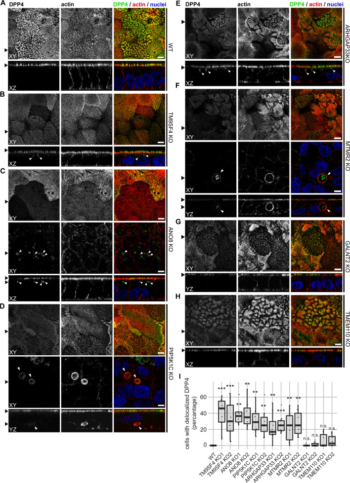
Figure 8.

Figure 8—figure supplement 1. Confocal micrographs of aminopeptidase N (APN) and actin immunofluorescence staining from wildtype (WT) and respective knockout (KO) cell lines.

Figure 8—figure supplement 1.

(A) APN localization is restricted to the actin-rich microvillus brush-border in WT cells. (B–D) APN localization is restricted to the actin-rich microvillus brush-border in TM9SF4-, PIP5K1C-, and ANO8-KO cells. Additionally, intracellular actin-positive compartments are detected (white arrowheads). (D) ARHGAP33-KO cell lines display large, APN- and actin-positive, intracellular compartments (white arrowheads). (E) DPP4 mislocalizes to subapical compartments in ARHGAP33-KO cells (white arrowheads). (F) APN localization is restricted to the actin-rich microvillus brush-border in MTMR2-KO cells. Additionally, intracellular actin-positive compartments are detected (white arrowheads). (G, H) In GALNT2- and TMEM110-KO cells, DPP4 localizes strictly to the apical plasma membrane. (A–H) XY = top view of polarized monolayer; XZ/YZ = lateral view of polarized monolayer. Scale = 5 µm. (I) Delocalized APN in polarized CaCo2 cells was quantified (dot box plot, Mann–Whitney U test. ***p<0.005, n.s., not significant, n ≥ 100 cells per condition).
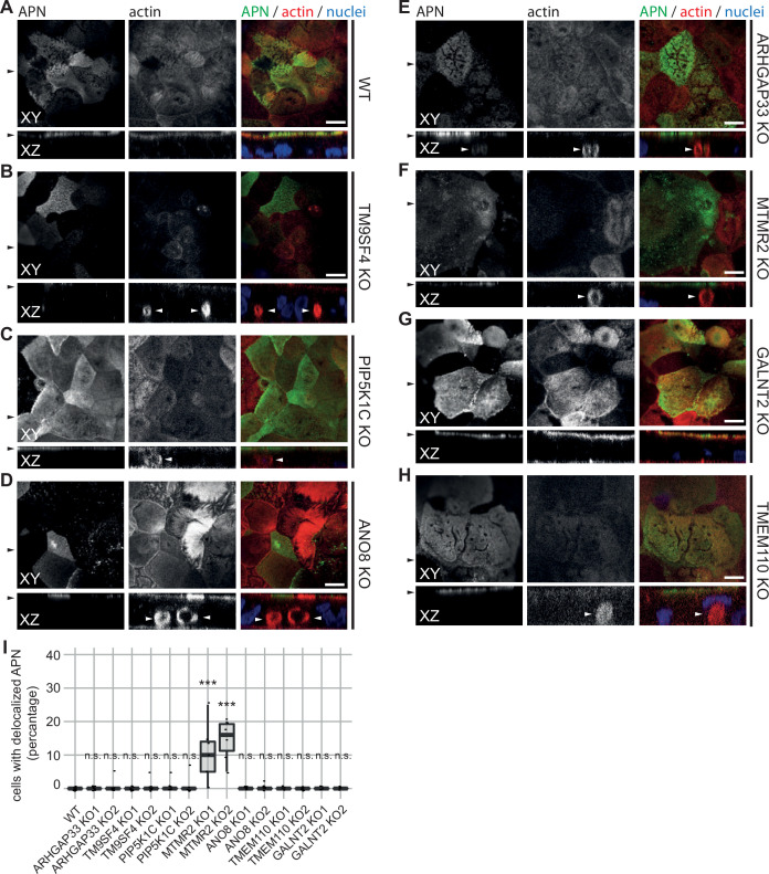
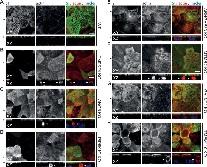
Figure 8—figure supplement 2. Confocal micrographs of surcase-isomaltase (SI) and actin immunofluorescence staining from wildtype (WT) and respective knockout (KO) cell lines.

Figure 8—figure supplement 2.

(A) SI localization is restricted to the actin-rich microvillus brush-border in WT cells. (B–H) KO of indicated genes does not alter the localization of SI localization is restricted to the actin-rich microvillus brush-border. XY = top view of polarized monolayer; XZ/YZ = lateral view of polarized monolayer. Scale = 5 µm.