
Figure 3.
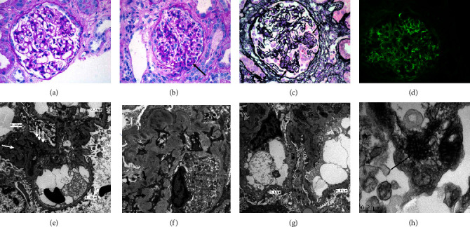
(a) Second biopsy: a glomerulus with no evidence of hypercellularity (PAS; ×400). (b) Second biopsy: a glomerulus with mild endocapillary proliferation (arrow) (PAS; ×400). (c) Secondary biopsy: a glomerulus with endocapillary proliferation in two segments (arrows) and, GBM breaks and crescent formation between the two segments (PAMS; ×400). (d) Secondary biopsy: deposition of granular immune complexes along the GBM and, to a lesser degree, within the mesangium (IF, IgG; ×400). (e) Second biopsy: thin GBM, intramembranous and para-mesangial electron dense deposits (arrows), and focal effacement of foot processes (EM; scale bar = 2 μ). (f) Second biopsy: intramembranous and mesangial electron dense deposits (arrows), and effacement of foot processes (EM; scale bar = 2 μ). (g) Second biopsy: thin GBM, intramembranous and para-mesangial electron dense deposits, endothelial cell swelling and loss of fenestration (EM; scale bar = 2 μ). (h) Tubulo-reticular inclusions within the endothelial cytoplasm (EM; scale bar = 22 μ).