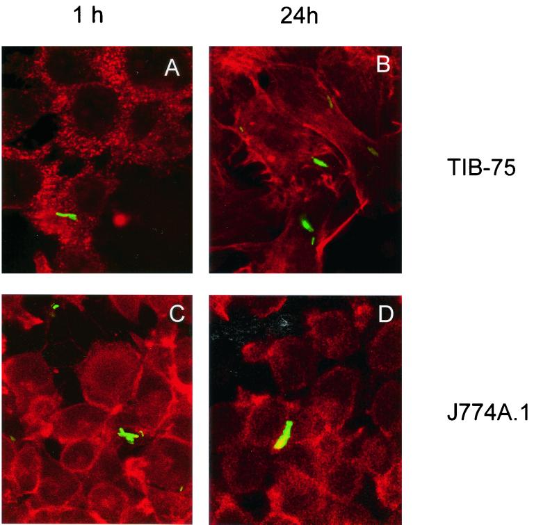
FIG. 2.
Confocal analysis of liver cell infection in vitro. TIB-75 mouse hepatocyte and J774A.1 mouse macrophage cell lines were infected with rBCG-GFP at an MOI of 10 for 1 h, resulting in an infection rate of 5 to 10% of the cells and analyzed after 1 h (A and C) and 24 h (B and D), respectively. For 24 h of infection, cultures were supplemented after 1 h of incubation with 25 mg of gentamicin sulfate per liter. Prior to analysis, cells were fixed with 4% PFA and stained with phalloidin-TRITC in order to visualize actin. The rBCG-GFP were localized within the cells by confocal laser microscopy. Shown are projections of eight confocal laser scans. Original magnification, ×1,000. Shown is one representative experiment of two similar ones.

