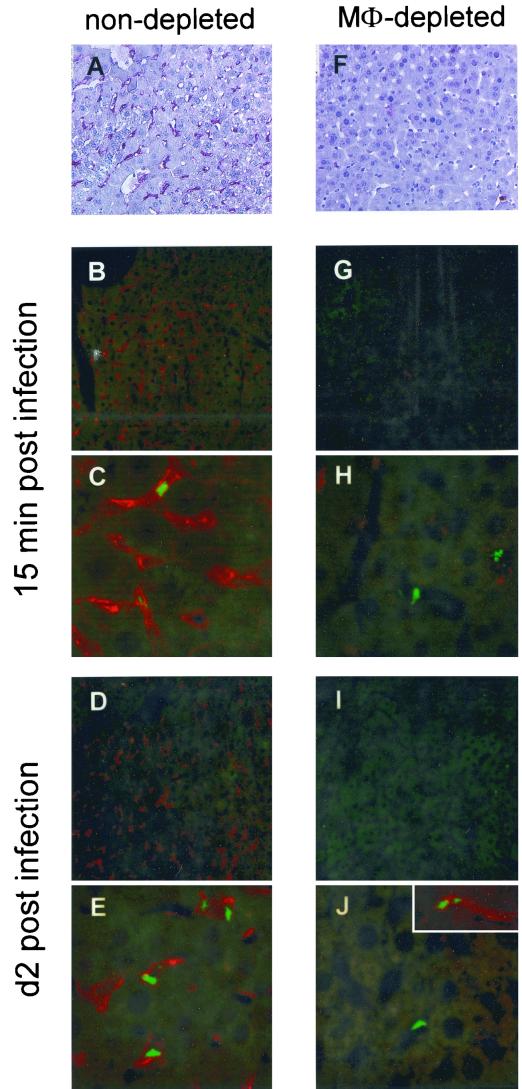
FIG. 3.
Confocal analysis of liver cell infection in vivo. C57BL/6 mice were either left untreated (A to E) or treated with 1.5 mg (F to J) of clodronate liposomes intraperitoneally at day −3. At day 0, mice were infected intravenously with 106 CFU of rBCG-GFP. At 15 min and 2 days postinfection, liver sections were stained with the rat MAb F4/80 and the secondary PAb goat anti-rat alkaline phosphatase (A and F) or PAb goat anti-rat Cy3.18 (B to E and G to J) in order to detect liver macrophages. Clusters of rBCG-GFP were detected inside the liver macrophages by confocal laser microscopy. Original magnification, ×200 (A, B, D, F, G, and I) and ×630 (C, E, H, and J). Shown is one representative experiment of two similar ones.

