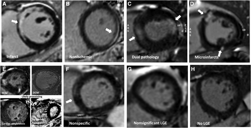
Figure 2.
Patterns of myocardial scar (late gadolinium enhancement). Patterns of late gadolinium enhancement (LGE; in parentheses, the features of each). A, Infarct (bright, subendocardial, or territorial). B, Nonischemic (mid-myocardial, less bright, or more diffuse). C, Dual pathology (both A and B). D, Microinfarcts (bright spots [eg, approximately 1 gram] of LGE often but not exclusively subendocardial and potentially in >1 territory). E, Chronic, likely preexisting disease (only 4 cases total; top left: HCM; top right: dilated cardiomyopathy [DCM]; bottom left: amyloidosis; bottom right: pulmonary hypertension). F, Nonspecific (unequivocal LGE that cannot be considered normal and has insufficient volume to assign with certainty to any other category). G, Nonsignificant LGE (minor right ventricle insertion point LGE alone; trabecular LGE alone; or septal perforator LGE alone, which can be considered normal variant). H, No LGE. Other examples are shown in Figures S1 and S2.

