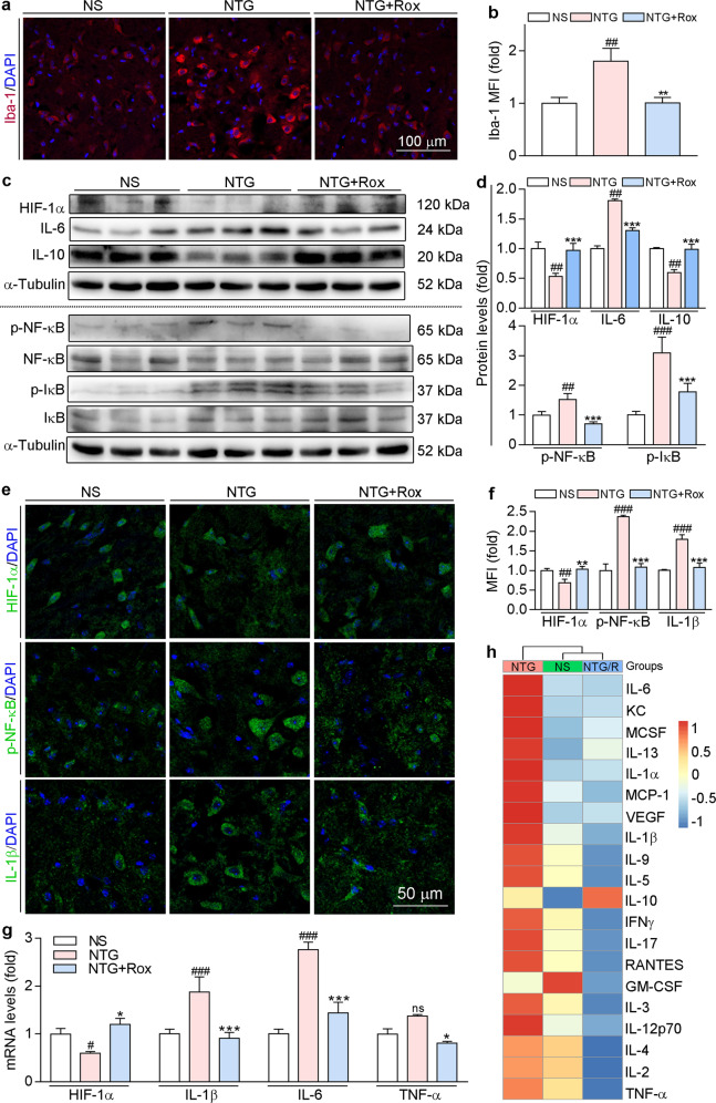
Fig. 7. Acute treatment with roxadustat ameliorates NTG-induced neuroinflammation and systemic inflammation.
Brain tissues and serum collected from mice used in Fig. 5 were conducted the following assays. a and b TNC frozen sections were used to determine Iba-1 protein expression by immunofluorescence staining with quantitation of MFI. c and d Protein expression levels of HIF-1α, IL-6, IL-10, NF-κB, p-NF-κB, IκB, and p-IκB in TNC were determined by Western blot with quantitative analysis of band intensity. e and f TNC frozen sections were used to determine HIF-1α, p-NF-κB, and IL-1β protein expression by immunofluorescence staining with quantitation of MFI. g mRNA levels of HIF-1α, IL-1β, IL-6, and TNF-α in TNC were determined by qRT-PCR. h Serum proinflammatory cytokines were analyzed by RayBio® Label-Based Mouse Antibody Arrays according to the manufacturer’s instructions. #P < 0.05; ##P < 0.01; ###P < 0.001 vs. NS group; *P < 0.05; **P < 0.01; ***P < 0.001 vs. NTG group (n = 3); ns: not significantly different, Rox: roxadustat.

