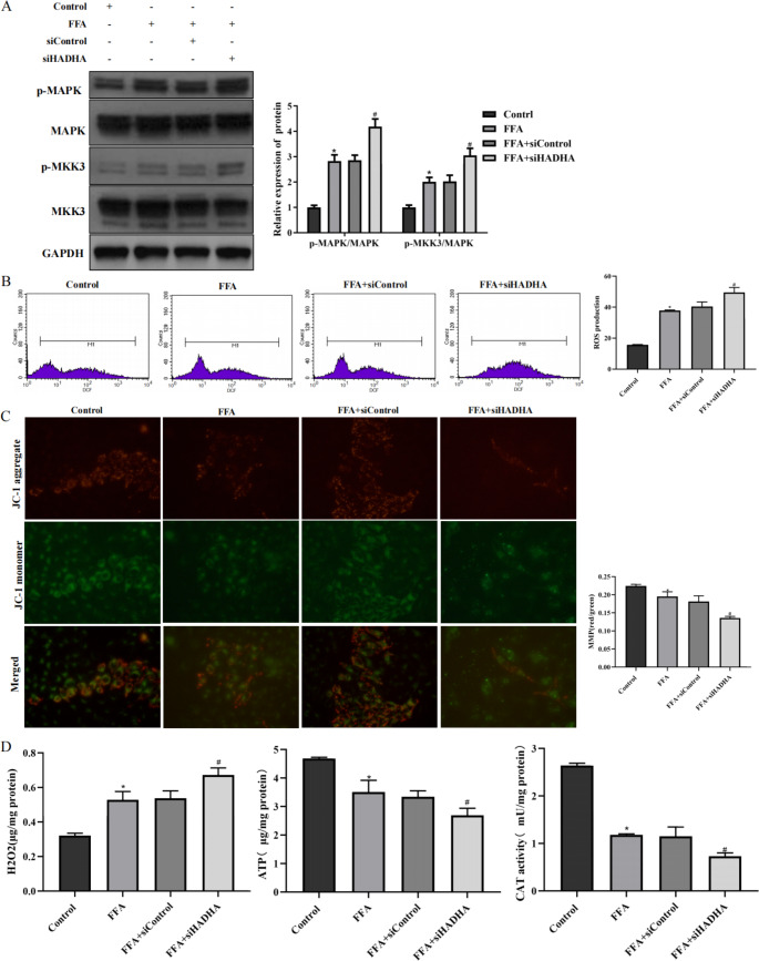
Fig. 3.
Inhibition of HADHA accelerated mitochondrial dysfunction and oxidative stress in L02 cells A: L02 cells were transfected with siHADHA or siControl for 24 h and then treated with 1 mM FFA for 24 h. The proteins p-MAPK, MAPK, MKK3 and p-MKK3 were examined by western blotting. B: DCF fluorescence staining was used to examine ROS production. C: A JC-1 assay was used to examine MMP (400×). D: The levels of H2O2, ATP, and CAT in L02 cells were examined by biochemical tests. *P < 0.05 compared with the control group; #P < 0.05 compared with the FFA + siControl group

