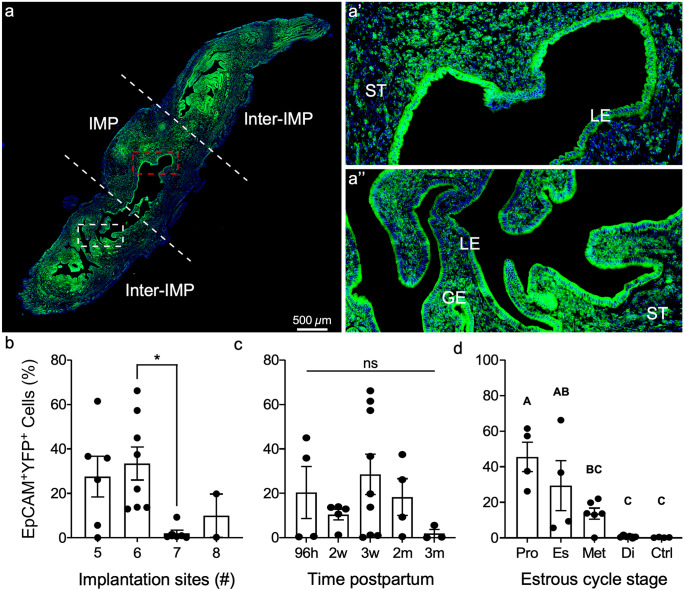
Fig. 2.
Mesenchymal-derived (MD) epithelial cells fluctuate across the estrous cycle in postpartum uteri. a Representative image of direct YFP fluorescence in a longitudinal section of a uterine horn following pregnancy and endometrial repair in Amhr2-Cre; Rosa-EYFP female mice. The implantation site (IMP) is demarcated by white dashed lines and is flanked by inter-implantation sites (Inter-IMP). a’ Magnified image of red-boxed area in (a) showing YFP+ (MD) and YFP− (non-MD) epithelial cells in the IMP site. a’’ Magnified image of white-boxed area in a showing YFP+ (MD) and YFP− (non-MD) epithelial cells in the inter-IMP site. The percentages of EpCAM+YFP.+ (MD-epithelial cells) analyzed by flow cytometry were quantified and graphed by the number if implantation sites (b), time postpartum (c), and stage of the estrous cycle (d). Statistical analyses were performed by one-way ANOVA with significance at P < 0.05, indicated by * or different letters. ns, not significant; ST, stroma; LE, luminal epithelium; GE, glandular epithelium; EpCAM, epithelial cell adhesion molecule; YFP, yellow fluorescent protein; Pro, proestrus; Es, estrus; Met, metestrus; Di, diestrus; Ctrl, control (no cre)

