Fig. 5.
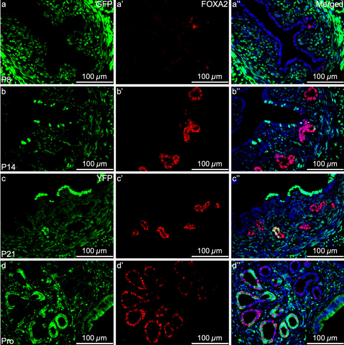
Mesenchymal-derived (MD) glandular epithelial cells express FOXA2. Representative images of uterine cross sections from Amhr2-Cre; Rosa26-tTA; H2B-GFP mice at postnatal day (P) 8 (a–a’’), P14 (b–b’’), P21 (c–c’’), and from Amhr2-Cre; Rosa-EYFP mice in proestrus (Pro) (d–d’’). (a, b, c, d) direct GFP/YFP expression in mesenchymal cells an MD-epithelial cells. (a’, b’, c’, d’) FOXA2 expression (red) by immunofluorescence, restricted to the glandular epithelium. (a’’, b’’, c’’, d’’) Merged images of the first two panels with nuclear DAPI staining (blue). FOXA2, Forkhead Box A2

