Fig. 6.
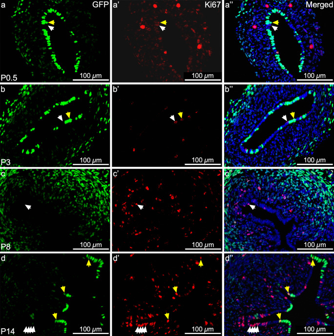
Mesenchymal-derived (MD) epithelial cells differentially contribute to epithelial remodeling by proliferation during postnatal uterine maturation. Representative images of uterine cross sections from Amhr2-Cre; Rosa26-tTA; H2B-GFP mice at postnatal day (P) 0.5 (a–a’’), P3 (b–b’’), P8 (c–c’’), and P14 (d–d’’). (a, b, c, d) Direct GFP expression in mesenchymal cells and MD-epithelial cells. (a’, b’, c’, d’) Ki67 expression (red) by immunofluorescence, indicating cells that proliferated. (a’’, b’’, c’’, d’’) Merged images of the first two panels with nuclear DAPI staining (blue). White arrow heads indicate GFP− (non-MD) epithelial cells that expressed Ki67. Yellow arrow heads indicate GFP.+ (MD) epithelial cells that expressed Ki67

